Figure 2.

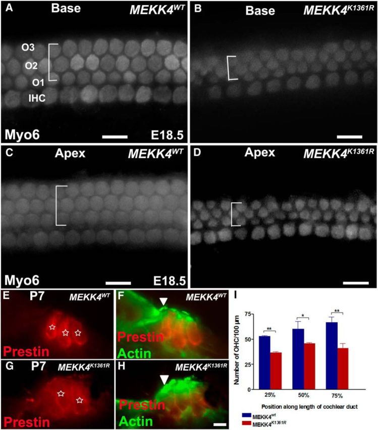
Disruption of MEKK4 pathway causes significant reduction in cells that differentiate into OHCs. A–D, Whole-mount surface views of the base (A, B) and apex (C, D) of a WT (A, C) and MEKK4K1361R mutant cochlea (B, D) at E18.5 immunolabeled with Myo6. In the WT cochlea, normal patterning of one row of IHCs and three rows of OHCs is present. However, in the mutants, the IHCs are unaffected; there are only two rows of OHCs with intermittent third row present in both basal and apical regions of the cochlear duct. The width of the lateral compartment of the sensory epithelium, as indicated by brackets, is diminished in the mutant cochlea compared with WT controls. E–H, Cross-sections of WT (E, F) and MEKK4K1361R cochleae (G, H) immunolabeled with Prestin (red) and actin (green) show that HCs (asterisks), still present at P7, are disorganized with less distinct separation between IHCs (F, H, arrowhead) and OHCs and closely packed OHCs in mutant cochlea as visualized by anti-Prestin (red). I, Quantification of HC counts shows significant decrease in the number of OHCs in homozygous MEKK4K1361R mutant cochlea, relative to MEKK4WT controls at distances 25%, 50%, and 75% from basal regions of the cochlear duct (n = 3). p < 0.001. Error bars indicate SEM. O1–O3, three rows of OHCs. Scale bars: (in D) A–D, 20 μm; (in H) E–H, 10 μm. * **
