Figure 3.
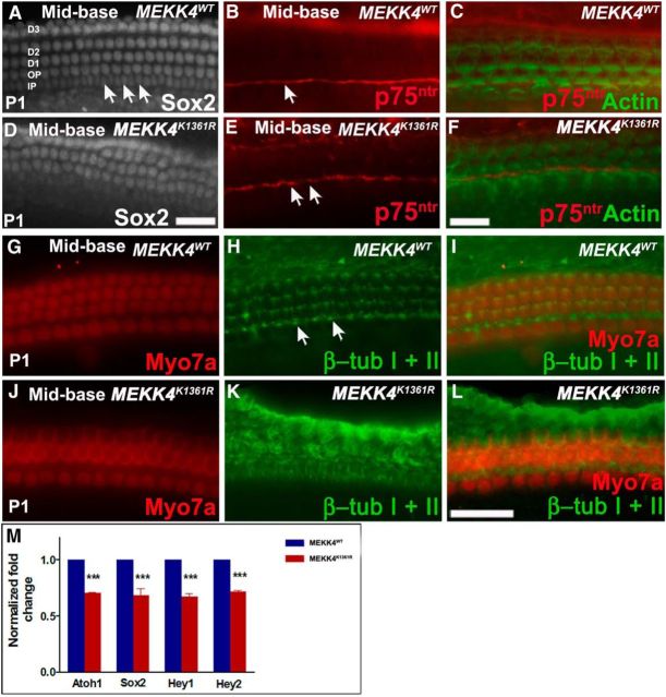
Supporting cell defects in MEKK4K1361R mutants. A–F, Whole-mount surface views of the mid-basal region of WT and mutant cochlea at P1 immunolabeled with Sox2 (A, D) or p75ntr (red) and actin (green) (B, C, E, F). A, In the WT, at least six rows of Sox2-positive supporting cells, including nuclei of inner and outer pillars, inner phalangeal cells, and three rows of Deiters' cells, are observed, which are arranged in organized rows. However, in mutants (D–F), disorganization of supporting cells is observed, and three rows of Deiters' cell nuclei are not discernible. Labeling of pillar cells is seen in the mutant organ of Corti with p75ntr comparable with WT control littermates (B, E, arrows) (B, C, E, F); however, the normal elongated shape of the nuclei of inner pillar cells (A, arrows) is not clearly seen, suggesting disruption of their morphological development. G–L, Labeling with β-tubulin I + β-tubulin II (green) reveals normal morphology of microtubule bundles within inner and outer pillar cell heads (arrows) and throughout the Deiters' cell rows in controls (G–I). J–L, In mutants, although microtubule bundles can be seen within inner pillar cells, microtubule bundle organization within outer pillar cells and Deiters' cells are indistinguishable. M, Relative expression levels of Atoh1, Sox2, Hey1, and Hey2 are significantly lower in mutant cochleae compared with controls (n = 3). p < 0.001. Error bars indicate SEM. D1–D3, Deiters' cell rows; OP, outer pillar cell row; IP, inner pillar cell row. Scale bars: A, D (in D), 20 μm; B, C, E, F (in F), 20 μm; G–L (in L), 20 μm.

