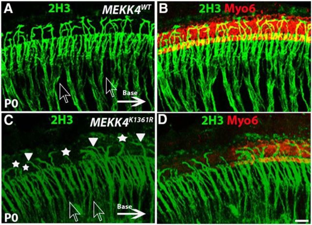
Figure 7.
Disruption of spiral ganglion neuron patterning in MEKK4K1361R mutants. Immunolabeling for Myo6 (red) and neurofilament 2H3 (green) in the mid-basal region of a WT (A, B) and mutant (C, D) cochlea at P0. In the WT cochlea, most of the afferent Type I fibers fasciculate to form bundles within otic mesenchyme (shown by clear arrows), before terminating on IHCs, whereas Type II afferent fibers, which constitute only 5% of afferent neurons, extend laterally to innervate OHCs and form outer spiral bundles before they turn toward the base (indicated by solid arrow) to form synapses with more than one OHC. C, In the mutants, disorganization of fibers from Type I neurons is quite evident and does not form bundles as indicated by clear arrows. Although most of these fibers terminate at IHC region, a smaller percentage of these fibers crossed the pillar cell region to reach OHCs compared with that in controls. Moreover, the fibers in the OHC region do not seem to turn toward the base (solid arrowheads); and instead, some fibers project laterally into the lesser epithelial ridge (asterisks). Scale bar: (in D) A–D, 20 μm.

