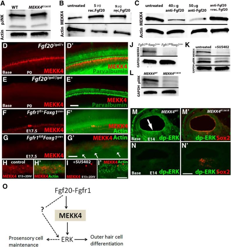
Figure 8.

MEKK4 function in the inner ear is independent of JNK activity and that its expression is activated by FGF20-FGFR1 signaling. A, Protein harvested from cochleae at E16.5 and blotted with anti-phospho-JNK show no alteration in its activity between control and MEKK4K1361R mutant samples. Data shown are representative results from three independent experiments. B, C, Immunoblot analysis of proteins harvested from WT and mutant cochlear explants treated with recombinant Fgf20 (B) or function-blocking antibody (C) at E13 show stimulation (B) or inhibition (C) of MEKK4 expression, respectively. The inhibition of MEKK4 expression in cochlear explants cultured in the presence of anti-Fgf20 was rescued by the addition of recombinant Fgf20 protein for 2DIV (C). D–E′, Immunolabeling of Fgf20βgal/+ heterozygote control (D, D′) and Fgf20βgal/βgal homozygous mutant (E, E′) inner ears at P0 using anti-MEKK4 (red) and anti-parvalbumin (green) demonstrates normal expression of MEKK4 in the cochlear duct; however, its expression is downregulated in Fgf20βgal/βgal cochleae. F–G′, Immunolabeling of Fgfr1fl/+foxg1cre+ (F, F′) and Fgfr1fl/flfoxg1cre+ (G, G′) cochleae at E17.5 using anti-MEKK4 (red) and actin (green) shows normal distribution of MEKK4 expression in controls, whereas mutant cochlea display patchy HCs (G′, arrows) and a complete absence of MEKK4 expression. H–I′, Control DMSO-treated (H, H′) and SU5402-treated (I, I′) cochlear cultures established at E13 and after 2 DIV immunolabeled using anti-MEKK4 (red) and actin (green) show normal expression of MEKK4 throughout the sensory epithelial cells in control cochleae, although its expression is completely lost in treated cochleae. J, K, Immunoblot-based assays show complete absence of MEKK4 expression within Fgfr1fl/flfoxg1cre+ mutant cochlea at E13 (J). In addition, E13 cochlear cultures treated with SU5402 for 12 h show rapid downregulation of MEKK4 and pERK levels (K). L–N′, Phospho-ERK levels (green) were also significantly downregulated in MEKK4K1361R compared with controls in the developing cochlear duct as shown by immunoblot (L) and immunolabeling (M–N′) (arrow in M indicates sensory patch) assays at E13 and E14 stages, respectively. O, Summary of the molecular signaling modules that control sensory cell differentiation during inner ear morphogenesis. FGF20 signals are transduced through FGFRs, most likely FGFR1, to MEKK4 and ERK to regulate differentiation of OHCs, whereas FGF signaling activation via yet unknown MAPK molecule, possibly another MEKK, controls cochlear progenitor cell population. Solid arrows indicate direct interaction between molecular components. Dotted line and arrows indicate hypothetical direct regulation. Scale bars: (in E′) D–E′, 20 μm; (in G′) F–G′, 20 μm; (in I′) H–I′, 20 μm; (in N′) M–N′, 10 μm.
