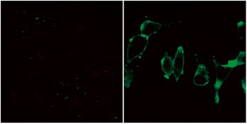
Fig. 3.

Natural expression of TLR4 in fibroblast. Cells were observed by confocal microscopy. Left image shows the negative control (rabbit anti-mouse TLR4 antibody was not added), with no cell stained by FITC, and cell outline only dimly seen. Right image shows spindle-shaped fibroblasts, with cell membrane staining showing that TLR4 was widely expressed and uniformly distributed on cell membrane. Magnification: ×560
