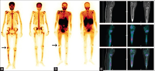
Figure 1.
Tc99m methylene diphosphonate bone scan (a) showing multiple foci of increased uptake in the skull bones, mandible, axial and appendicular skeleton with focally increased uptake in the upper and lower ends of both femurs, both tibia, both shoulder joints and forearm bones. 99mTc-MIBI WBS (b) showing increased uptake in the mandible, upper and lower ends of the right femur, both tibial bones, and around the shoulder joints, similar to that of the bone scan and SPECT/CT (c) of the lower limb that showed MIBI avid lytic-cystic lesions in the lower end of the right femur and midshaft of both tibial bones

