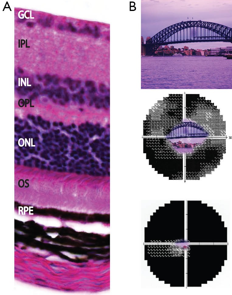
Figure 1.

Retinal layers and clinical impact of retinal dystrophy. (A) Expansion of retinal region with histology section showing normal retinal layers of the mouse, which has close homology with histology of the human eye. The ONL contains the nuclei of the photoreceptors, and the OS layer contains the outer segments of the photoreceptors; (B) progressive visual field loss as experienced by retinitis pigmentosa patients. Upper image shows normal full visual field, middle image shows constricted field and lower image shows almost no central vision remaining. GCL, ganglion cell layer; IPL, inner plexiform layer; INL, inner nuclear layer; OPL, outer plexiform layer; ONL, outer nuclear layer; OS, outer segments of photoreceptors; RPE, retinal pigment epithelium.
