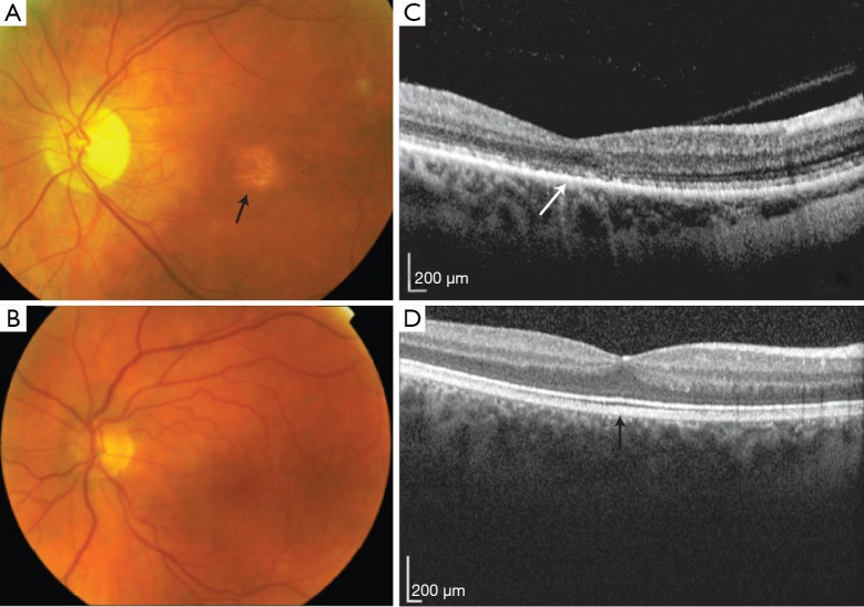
Figure 3.
Cone dystrophy: fundal images. (A,B) Wide field fundus photography illustrating retinal features of cone dystrophy, showing macular atrophy in (A) (arrow), compared with the normal macular appearance present in (B); (C,D) OCT in cone dystrophy illustrates the loss of the foveal photoreceptor outer segments in (C), compared with normal in (D) (arrows). OCT, optical coherence tomography.

