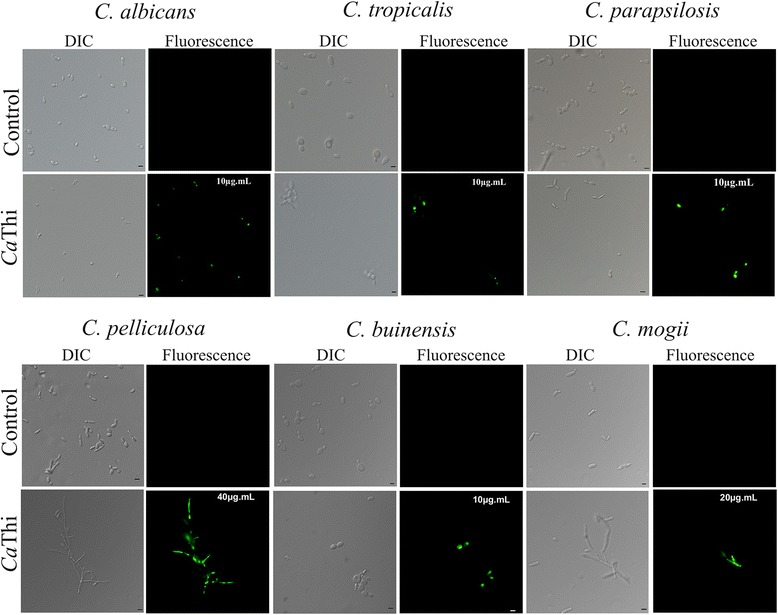
Fig. 2.

Membrane permeabilization assay. Photomicrography of different yeast cells after membrane permeabilization assay by fluorescence microscopy using the fluorescent probe Sytox green. Cells were treated with CaThi for 24 h and then assayed for membrane permeabilization. Control cells were treated only with probe Sytox green. Bars 5 μm
