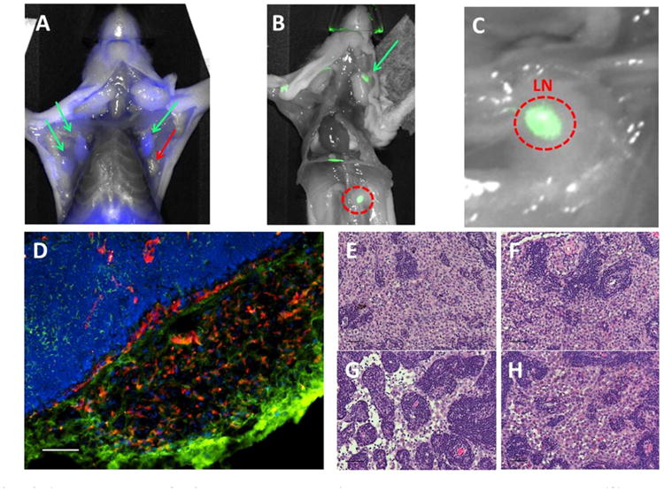
Fig. 8.

PSMA-expressing JHU-LNCaP-SM subcutaneous xenografts spontaneously metastasize to lymph nodes. (A) Ex vivo PSMA-targeted optical imaging of lymph nodes from selected mice that exhibited spontaneous metastasis from subcutaneous JHU-LNCaP-SM primary xenografts where green arrows show PSMA positive axillary lymph nodes and red is a negative node. (B and C) The red circle is a positive renal lymph node and also shown is a superficial cervical lymph node denoted by a green arrow. (D) Frozen sections of lymph node metastasis were analyzed by immunofluorescence for PSMA and CD31 expression and confirm the presence of PSMA-expressing cells within a vascular cage at the edge of packed lymphatic cells (scale bar = 100 μm). (E–H) H&E staining of various PSMA positive nodes confirming the presence of tumor cells (scale bar = 50μm).
