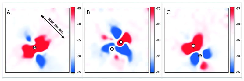
Figure14_7.
Virtual electrode patterns optically recorded from a 4 × 4-mm area of the rabbit anterior epicardium during unipolar and bipolar stimulation. A: conventional “dogbone”-shaped virtual electrode polarizations (VEP) during unipolar cathodal stimulation. B: VEP during bipolar stimulation with a pacing dipole placed perpendicular to myocardial fibers. C: results of bipolar stimulation with electrodes along the fibers. The interelectrode distance was 0.8 mm. Images were collected in the middle of a 2-ms diastolic stimulus. This figure is modified from Nikolski et al.[47]

