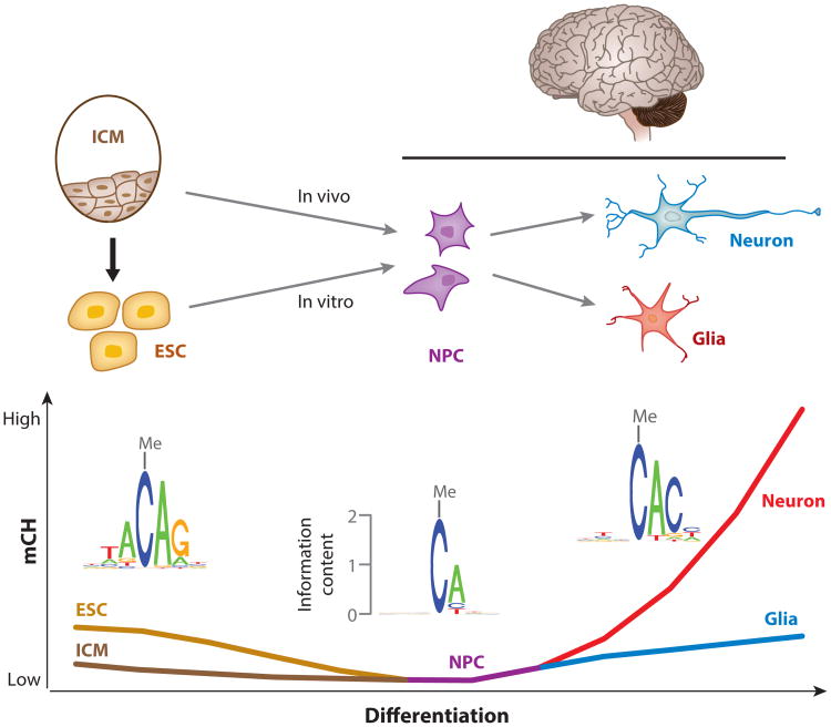
Figure 6.
Hypothesized reconfiguration of mCH patterns during neural differentiation. Abundant mCH is present in ESCs derived from ICM cells, which show low levels of mCH. During the in vitro differentiation process from ESCs to NPCs, non-CG demethylation takes place, and genome-wide mCH is lost in NPCs. When NPCs differentiate into neurons and glia, the genome-wide mCH could possibly be reestablished in in vitro differentiated neurons and glia, but methylation measurements to support this hypothesis are lacking. The absence of mCH in NPCs and the reestablishment of mCH in neurons and glia during in vivo neural differentiation are supported by methylation data (54), but whether mCH is absent in intermediate cells between ICM cells and NPCs is unclear. In addition, the fact that mCH occurs primarily at CAG sites in ESCs but at CAC sites in both neurons and glia implies that an interesting switch of mCH pathways occurs during neural differentiation. Abbreviations: ESC, embryonic stem cell; ICM, inner cell mass; mCH, non-CG methylation; NPC, neural progenitor cell.

