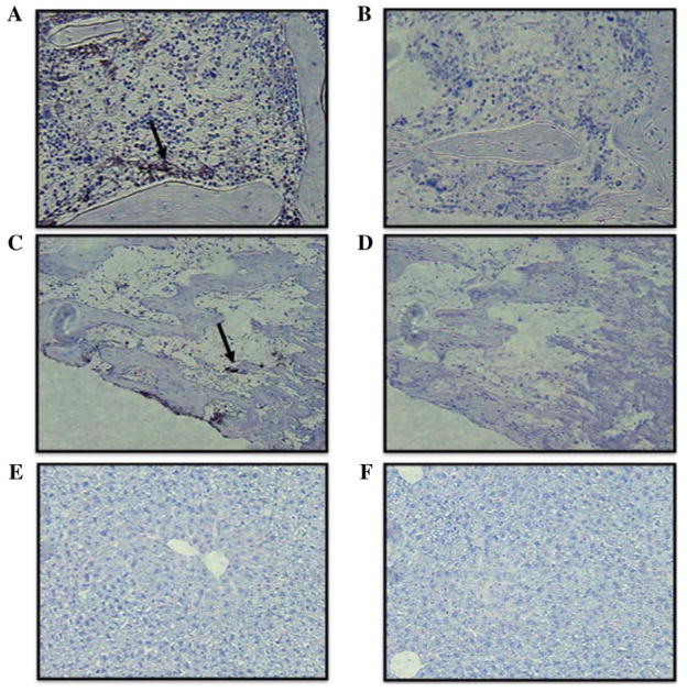
Fig. 5.
Immunochemical analysis of infused MSC–GFP (stained in black) (A) Distribution of infused GFP-expressing MSC in the bone marrow of the femur (arrow, 10x), and (B) control without antibody; (C) Distribution of infused GFP-expressing MSC in the bone marrow of the tibia (arrow, 10x), and (D) control without antibody; (E) Absence of GFPexpressing MSC in the liver (10x), and (F) control without antibody.

