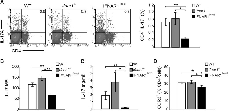
Figure 7. Selective T cell IFNAR signaling leads to a decreased Th17 response at the presymptomatic phase of EAE.
Splenocytes were isolated from WT, Ifnar1−/−, and IFNAR1Texcl mice, 10 d upon EAE induction. (A) Cells were stimulated with PMA/ionomycin/brefeldin A, and the frequency of IL- 17A-producing CD4+ T cells in the spleen was determined. The dot plots and bars represent IL-17A+ cells gated on total splenocytes. A representative dot plot per genotype is shown. (B) Expression levels of IL-17A by CD4+ T cells were evaluated by measuring MFI from (A) dot plots. (C) Splenocytes from all groups were restimulated ex vivo with MOG35–55 for 72 h, and the levels of secreted IL-17A were measured. (D) Frequency of CCR6+ splenocytes was determined (gated on the CD4+ compartment). All results are shown as means ± sem (n = 5 mice/genotype) and are representative from 2 independent experiments with similar results. *P < 0.05, **P < 0.01, ***P < 0.001.

