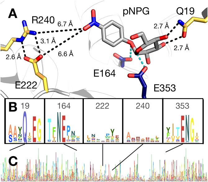
Fig 3. Active site model and conservation analysis of BglB.
(A) Docked model of pNPG in the active site of BglB showing established catalytic residues (navy) and a selection of residues mutated (gold). A multiple sequence alignment of the Pfam database’s collection of 1,554 family 1 glycoside hydrolases was made and the sequence logo for (B) selected regions around specific residues discussed in the text and (C) over the entire BglB coding sequence is represented. The height for each amino acid indicates the sequence conservation at that position.

