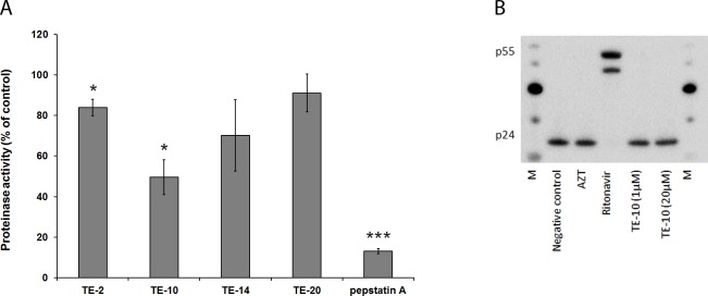
Fig 8. Inhibition of HIV-1 protease.
(A) Inhibition of HIV-1 protease activity: The HIV-1 protease (100 ng/well) and the compounds (25 μM concentration) or an inhibitor control (pepstatin A) were added to a flat-bottom 96-well plate. The plate was incubated for 15 minutes at 37°C and the provided protease substrate was added to all wells. The fluorescence was measured at Ex/Em = 490 nm/520 nm. The value for the vehicle control was set to 100% in each individual experiment and all other values were normalised to this reference value. The data are means of three independent experiments; the error bars represent the SEM. P-values are indicated if significant (*: p < 0.05; **: p < 0.01; ***: p < 0.001).(B) Inhibition of HIV-1 protease activity in viral particles: Persistently infected HuT-78/HIV-1 cells were incubated in the presence or absence of compound (3.7 μM AZT, 2.8 μM ritonavir or 1μM/20μM TE-10). After 43 h, virus particles in the cellular supernatants were harvested, lysed and subjected to Western blotting. P24 and its precursor p55 were detected using an anti-p24 antibody. Negative control: no compound added during incubation. M: MagicMark XP Western Protein Standard.

