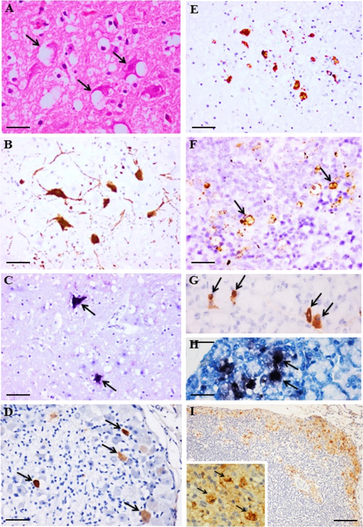
Fig 4. Pathological findings in CNS and non-CNS tissues from EV-A71 infected hamsters at day 4 post infection.
Vacuolation and degeneration in brainstem neurons (A, arrows) that also demonstrated viral antigens (B) and RNA (C, arrows). Viral antigens were also detected in dorsal root ganglion (D, arrows) and spinal cord anterior horn cells (E). Viral antigens in the spleen (F, arrows), salivary gland acinar cells (G, arrows), and lymph node (I and inset, arrows), and viral RNA in the salivary gland acinar cell (H, arrows) were observed. Stains: Hematoxylin and eosin (A), immunohistochemistry with 3, 3’ diaminobenzidinetetrahydrochloride chromogen/hematoxylin (B, D, E, F, G, I), and in situ hybridization with nitroblue tetrazolium/5-bromo-4-chloro-3-indolyl phosphate/hematoxylin (C, H). Original magnification: 10x objective (I), 20x objective (B-E), 40x objective (A, F, G, H, I inset). Scale bars: 50μm (I), 30μm (B-E), 15μm (A, F, G, H, I inset).

