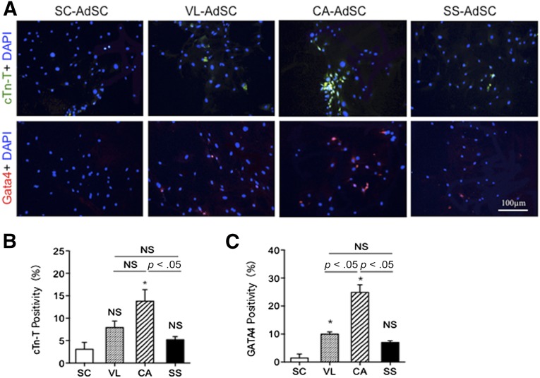
Figure 4.
Cardiomyocyte differentiation in AdSCs from four different adipose tissues by fluorescent immunocytochemistry. (A): To assess which source for AdSCs could differentiate into cardiomyocytes, the cells were stained with anti-cTnT (green) and anti-GATA4 (red) antibodies. Nuclei were stained with DAPI (blue). (B, C): The rate of cTnT-positive (B) and GATA4-positive (C) cells was compared among AdSCs from four different adipose tissues. ∗, p < .05; NS, not significant vs. SC. All experiments were performed in triplicate and statistically analyzed. Abbreviations: AdSCs, adipose-derived stem cells; CA, cardiac brown adipose tissue; cTn-T, cardiac troponin T; DAPI, 4′,6-diamidino-2-phenylindole; NS, not significant; SC, subcutaneous white adipose tissue; SS, subscapular brown adipose tissue; VL, visceral white adipose tissue.

