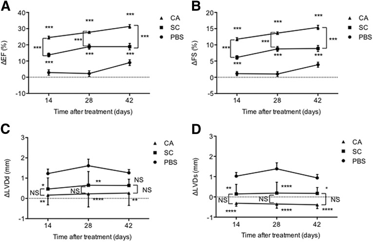
Figure 5.
Echocardiographic assessment of cardiac function after myocardial infarction with AdSC transfusion. The cardiac functional parameters in mice were assessed by echocardiography. The EF (A), FS (B), LVDd (C), and LVD (D) before and after treatment (14, 28, and 42 days) were measured, and the changes in each parameter (ΔEF, ΔFS, ΔLVDd, and ΔLVD) were statistically analyzed. ∗, p < .05; ∗∗, p < .01; ∗∗∗, p < .001; ∗∗∗∗, p < .0001; NS, not significant vs. PBS. Abbreviations: Δ, change; AdSCs, adipose-derived stem cells; CA, cardiac brown adipose tissue; EF, ejection fraction; FS, fractional shortening; LVD, left ventricular end-systolic dimension; LVDd, left ventricular end-diastolic dimension; NS, not significant; PBS, phosphate-buffered saline; SC, subcutaneous white adipose tissue.

