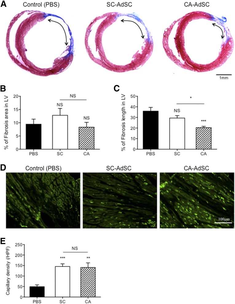
Figure 6.
Cardiac morphometric analysis after myocardial infarction with AdSC transfusion. (A): Mouse cardiac cross-sections of control group (PBS), SC-AdSC-treated group, and CA-AdSC-treated group through infarcted myocardium 42 days after surgery were assessed by Masson’s trichrome staining. The percentage of the mouse LV fibrotic area (B) and LV fibrosis length (C) were measured. (D, E): Mouse cardiac cross-sections were stained with an anti-BS-1 lectin antibody and the endothelial cell marker (green), and the capillary densities were compared among the PBS, SC-AdSC, and CA-AdSC groups. ∗, p < .05; ∗∗, p < .01; ∗∗∗, p < .001; NS, not significant vs. PBS. Abbreviations: AdSCs, adipose-derived stem cells; CA, cardiac brown adipose tissue and CA-AdSC group; HPF, high power field; LV, left ventricle; NS, not significant; PBS, phosphate-buffered saline; SC, subcutaneous white adipose tissue and SC-AdSC group.

