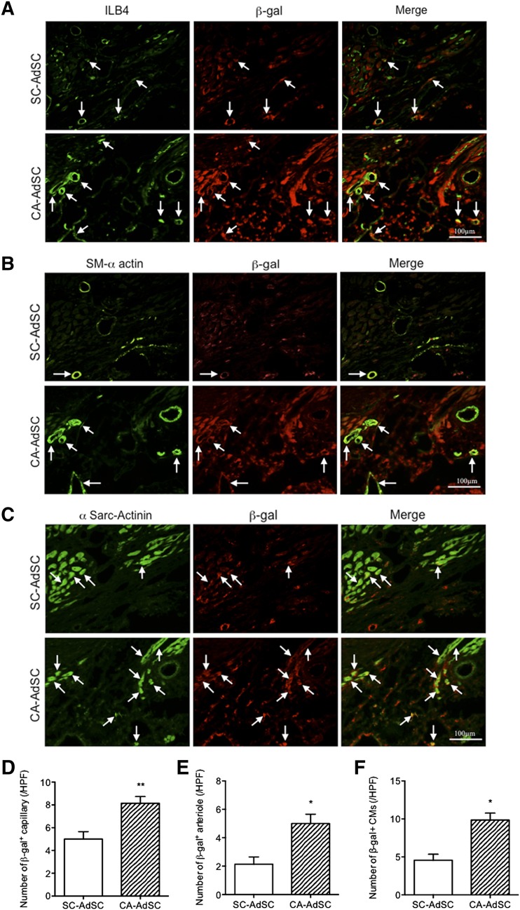
Figure 7.
Immunohistological assessment of ischemic myocardium with recruited β-gal expressing AdSCs. Mouse cardiac cross-sections of SC-AdSC-treated group and CA-AdSC-treated group through infarcted myocardium 42 days after surgery were stained with β-gal costained with ILB4, an endothelial cell marker (green) (A), SM-α-actin, a smooth muscle cell marker (green) (B), and a cardiac myocyte marker (green) (C). Arrows indicate the positive cells, where ILB4 (A), SM-α actin (B), and α-Sarc-actinin (C) was costained with β-gal. The double-positive cells for β-gal and ILB4, β-gal and SM-α actin, and β-gal and α-Sarc-actinin were quantified and expressed as the number of β-gal+ capillary (D), number of β-gal+ arteriole (E), and number of β-gal+ CMs (F), respectively. ∗, p < .05; ∗∗, p < .01 vs. SC-AdSCs. Abbreviations: α-Sarc-Actinin, α-sarcomeric actinin; AdSCs, adipose-derived stem cells; β-gal, β-galactosidase; CA, cardiac brown adipose tissue; HPF, high power field; ILB4, isolectin-B4; SC, subcutaneous white adipose tissue; SM-α actin, smooth muscle α-actin.

