Figure 1.
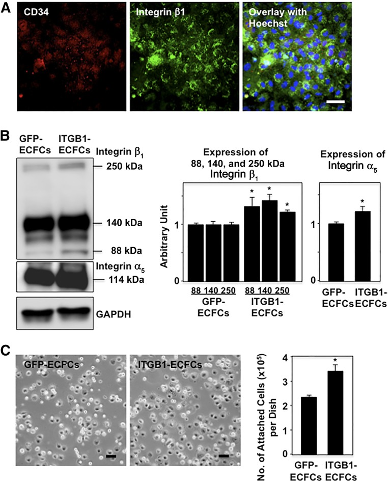
Genetically modified human ECFCs. (A): Confocal micrographs showing CD34 (red) and integrin β1 (green) immunostaining in human ECFCs transfected with integrin β1. Integrin β1 localized strongly at the cell surface. Scale bar = 50 μm. (B): Western blotting for integrins. In addition to integrin β1 (88 and 140 kDa), integrin α5 (114 kDa) was more strongly coexpressed in ITGB1-ECFCs than in GFP-ECFCs. The band at approximately 250 kDa suggests possible dimer formation between integrins β1 and α5 to form integrin α5β1, a fibronectin receptor. Graphs show densitometry for integrin β1 and integrin α5 (n = 3 each). (C): In vitro homing experiment for ECFCs. A significantly greater number of the cells from the ITGB1-ECFC group attached to the bottom of fibronectin-coated dishes compared with those from the GFP-ECFC group. Scale bars = 50 μm; ∗, p < .05 versus the GFP-ECFC group. Abbreviations: ECFCs, endothelial colony-forming cells; GAPDH, glyceraldehyde-3-phosphate dehydrogenase; GFP, green fluorescent protein; ITGB1, lentiviral vector encoding integrin β1.

