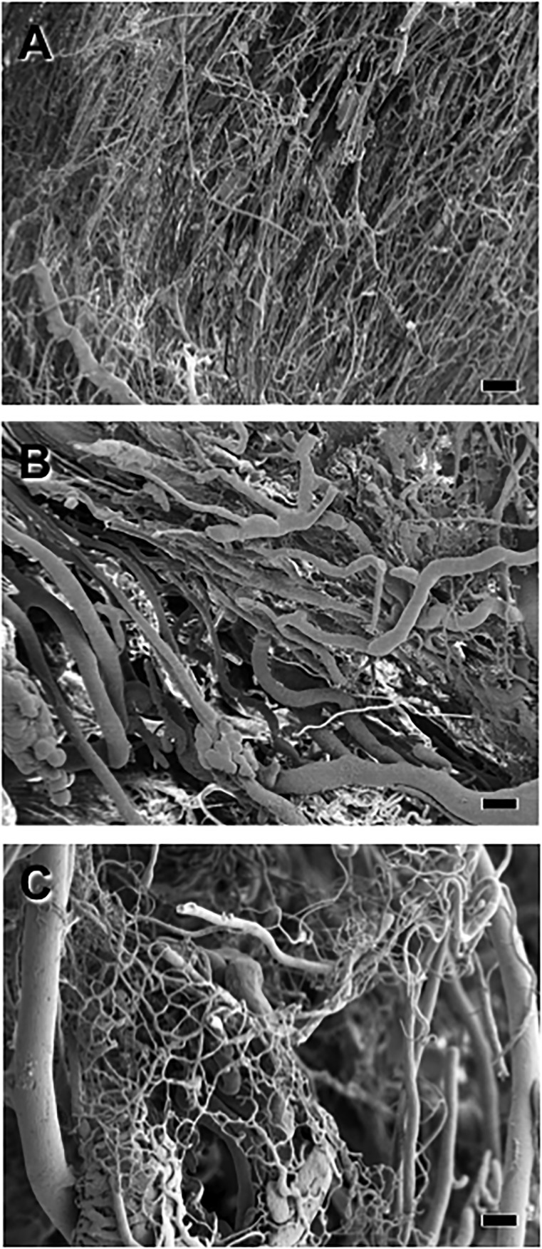
Figure 4.
Scanning electron photomicrographs of vascular casts of the hindlimbs 28 days after femoral artery ligation. (A): Vascular casts from a sham-operated mouse showing normal vascular profiles. (B): Vascular casts showing the less dense vasculature in the ischemic leg of a phosphate-buffered saline-treated mouse. (C): Vascular casts from the ischemic hindlimb of a mouse systemically treated with endothelial colony-forming cells overexpressing integrin β1. Note that the vasculature was substantially restored in this mouse. Scale bars = 10 μm.

