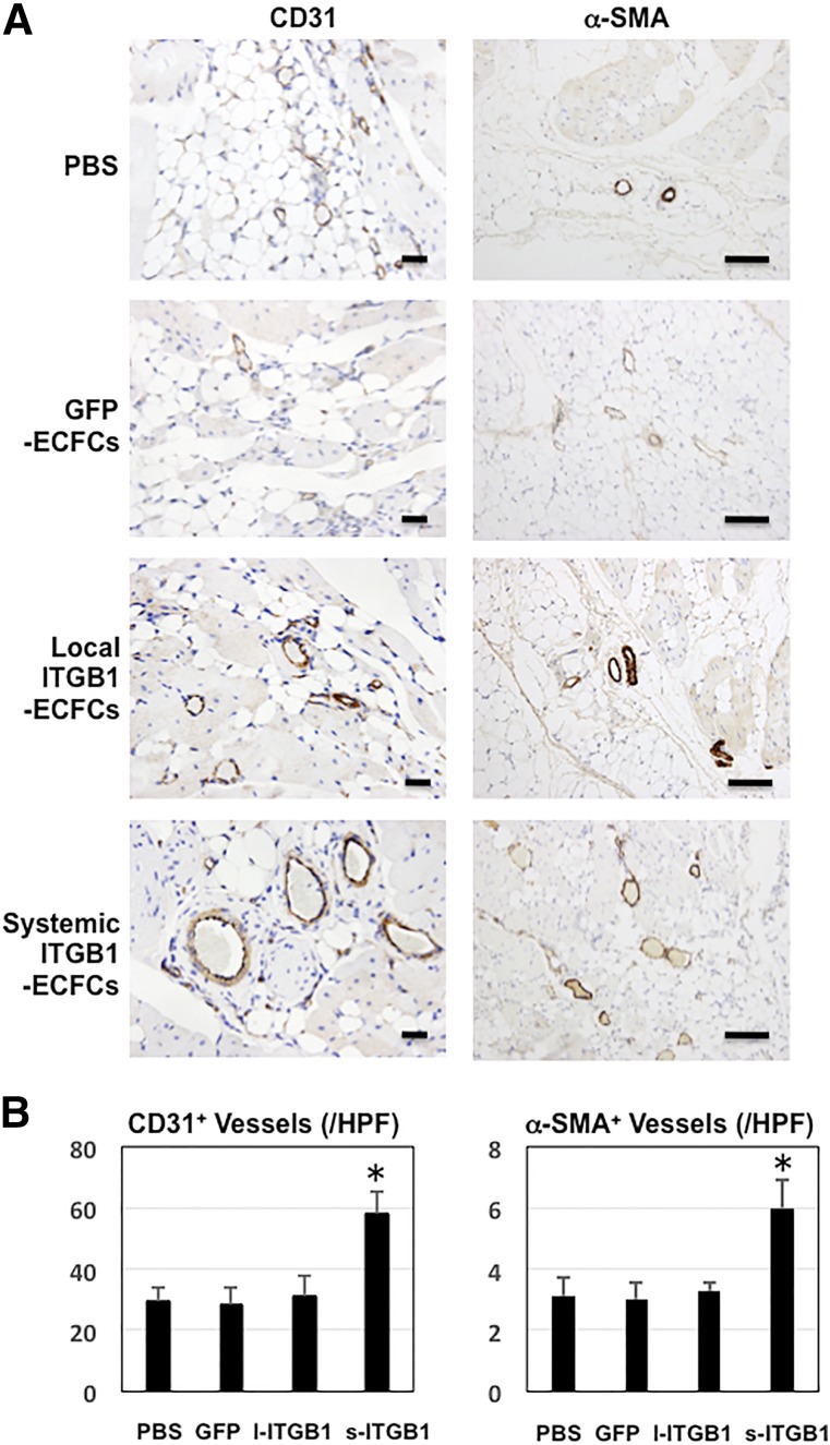
Figure 5.
Vascular immunohistochemistry. (A): Photomicrographs showing immunohistochemical staining of CD31- and α-SMA-positive lumens revealing total vessels (mainly capillaries) (left) and small arteries (right) in ischemic hindlimb tissues 28 days after surgery (n = 6 in each group), respectively. Scale bars = 50 μm. (B): Graphs showing densities of total vessels (left) and small arteries (right) in each group (n = 6 in each group). ∗, p < .05 versus the PBS, GFP-ECFC, and local ITGB1-ECFC groups. Abbreviations: α-SMA, α-smooth muscle actin; ECFC, endothelial colony-forming cell; GFP, green fluorescent protein; HPF, high-power fields; l, local; ITGB1, lentiviral vector encoding integrin β1; s, systemic.

