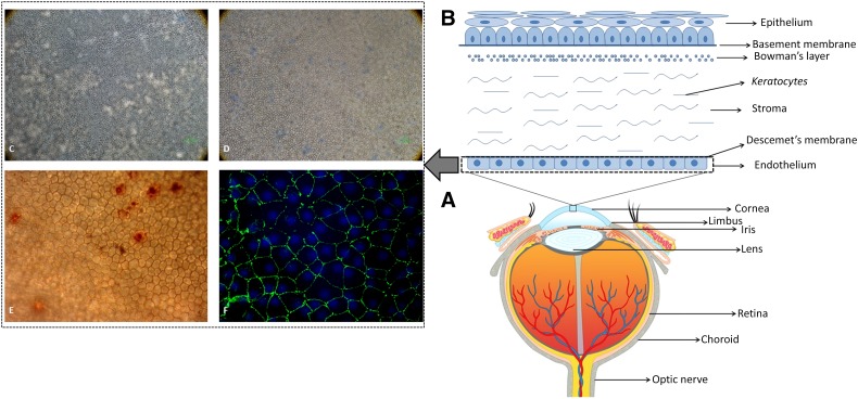
Figure 1.
Human eye, cornea, and endothelium. (A): Anatomy of human eye globe showing different parts of the eye. (B): Schematic representation and structure of human cornea showing specific layers of the tissue. (C): A normal human corneal endothelium seen under an inverted microscope at ×100 magnification. (D): Human corneal endothelium with high mortality rate observed using trypan blue staining at ×100 magnification. (E): Human corneal endothelium observed using alizarin red staining to check the hexagonality of the cells at ×200 magnification. (F): Human corneal endothelium expressing zonula occludens 1 marker observed under oil immersion magnification.

