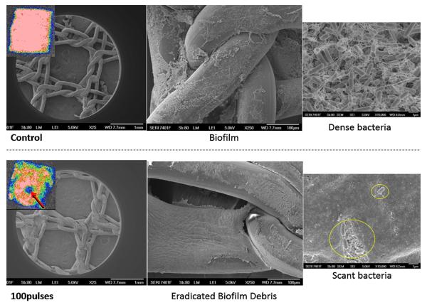
Figure 3.
Scanning Electron Microscopy confirmed our results. Control untreated infected mesh demonstrated thick biofilm wedged into mesh interstices. Dense bacteria revealed production of exopolysaccharide. After treatment with PEF using the conentric electrodes, the biofilm has been disrupted and debris is left behind. The few remaining scant rods displayed abnormal morphology and exopolysaccharide was not visible. The mesh was not damaged by PEF treatment.

