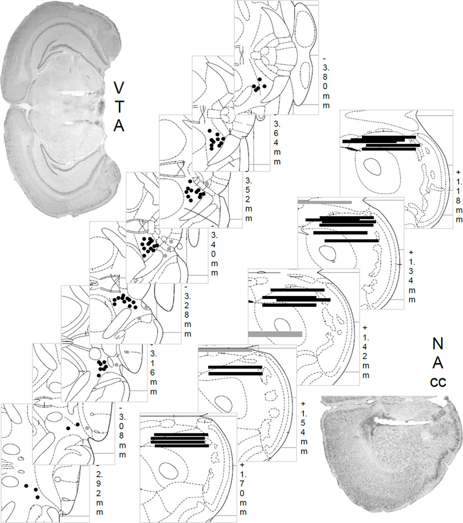
Fig 4.
Histological placements of guide cannula into the VTA for microinjection (black circles, n=62) or outside the VTA not included in analysis (grey circles, n=8). 1-mm microdialysis probes are shown into the NAc (black lines, n=21) or outside the NAc (grey lines, n=4). Representative photomicrographs for each brain site are shown.

