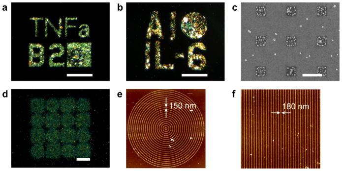
Figure 5.
Cytokine detection with a bound silver enhanced gold nanoparticles immunoassay on surface immobilized micro- and nano- patterns. Two cytokines, a) TNFα and b) IL-6 were detected from cell media of RAW 264.7 macrophages, scale bars = 35 μm. c) electron micrograph of anti-TNFα submicron patterns, scale bar = 1 μm. Anti-TNFα nanopatterns showing d) dark field micrograph cytokine detection immunoassay of circles and squares with nanometer line widths, scale bar = 20 μm, e) AFM of one of the circle patterns with 150 nm line widths, and f) AFM of one of the square patterns with 180 nm line widths indicated by the arrows.

