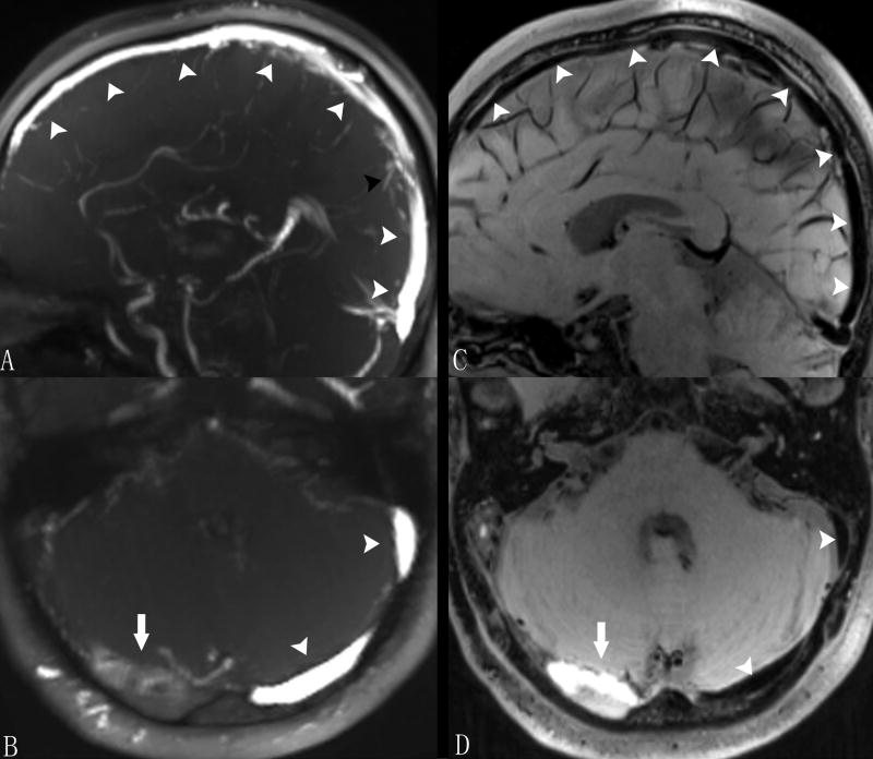
Figure 1.
MRBTI images of a 48-year-old male with sub-acute CVT. On TOF images, normal venous sinuses were depicted with bright venous flow signals (arrowheads in A and B) and minor flow defects (black arrowhead in A) observed in the superior sagittal sinus was not considered as thrombus by radiologists. With blood signal adequately suppressed on MRBTI, normal venous sinus were depicted as black area (arrowheads in C and D) and hyper-intense signal was found in the right transverse sinus suggesting a fresh thrombus (arrow in D). The thrombus was also confirmed on TOF (arrow in B).

