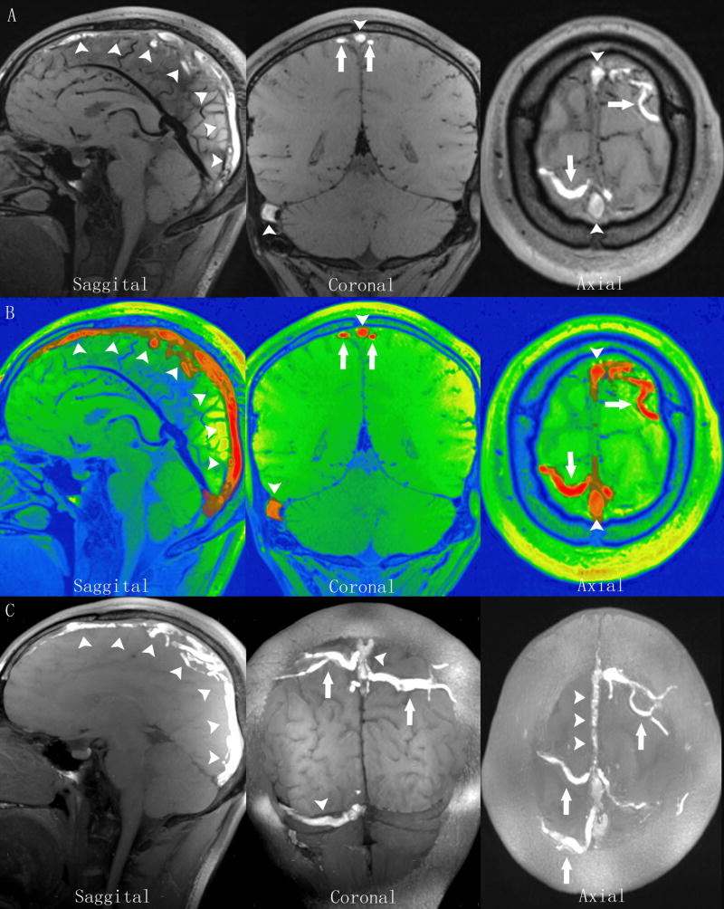
Figure 3.
MRBTI of a 27-year-old male patient with sub-acute CVT. A: MRBTI demonstrated hyper-intense signal intensity in the superior sagittal sinus (arrowheads), the right transverse and sigmoid sinuses (arrowheads), and the cortical veins (arrows) suggesting intraluminal thrombus formation. B: All thrombi semi-automatically outlined by software based on their high signal contrast were rendered with red color and volume was 21.5 cc. C: sagittal, coronal and axial sections of maximum intensity projection (MIP) reformations of MRBTI better depicted the thrombosed segments with hyper-intense signals.

