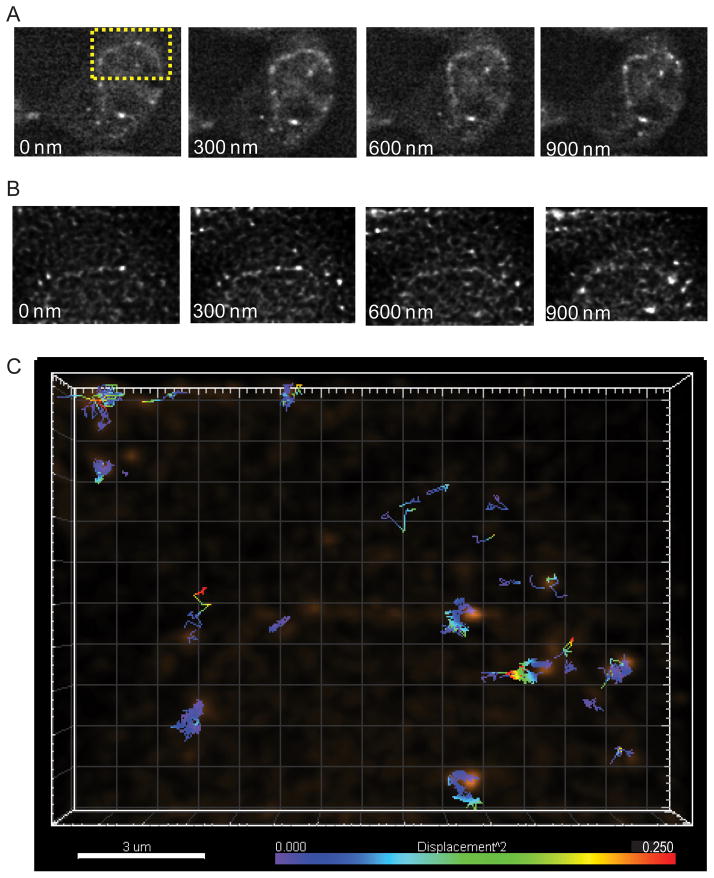
Fig. 2.
Multifocus imaging of HIV-1 genomic RNAs. (A) Multifocus fluorescence images of HIV-1 genomic RNAs (V1B-MS2) in HeLa MS2-NLS-mCherry cells. Images are 29.2 μm × 24.1 μm. (B) Multifocus images of HIV-1 genomic RNAs after deconvolution and alignment (images are the projection of the sum of intensity over 10 time points). Images are 14.7 μm × 10.4 μm. (C) A 3D multifocus image depicting individual tracks color-coded to represent the single step squared displacement of HIV-1 genomic RNAs.

