
Figure 2.
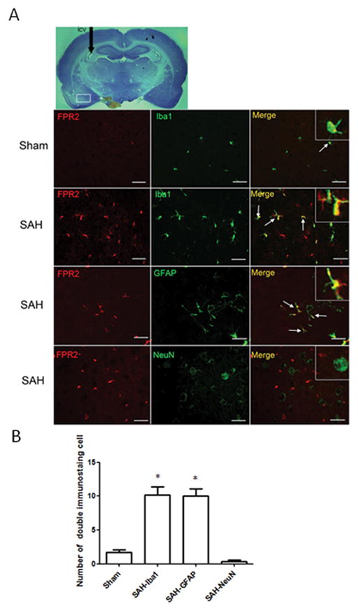
Immunostaining of FPR2 at 24 hours in Sham and SAH animals. (A) Double staining of FPR2 with Iba1, GFAP and NeuN. (B) Statistical analysis of the double staining cells. The expression of FPR2 is low in brain in sham animals, and SAH increased its expression in microglias and astrocytes, but not in neurons at 24 hours after SAH. n=3 for each group. *p<0.05 vs. Sham. Scale bar=30 μm.