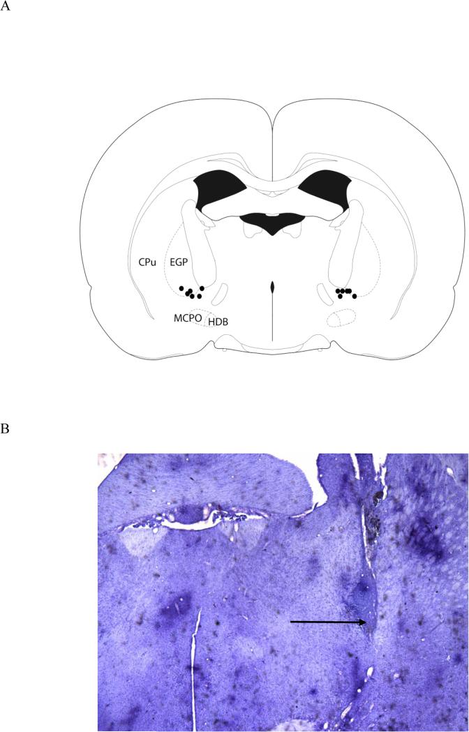
Figure 1.
The figure depicts the location of cannula placements for the six rats that were included in the analyses of attentional task performance (A). A photomicrograph, taken with a 2X objective, shows the cannula placement in one hemisphere of one animal, although all infusions were bilateral (B). The arrow indicates the infusion site. The section is approximately −1.3 mm from bregma.

