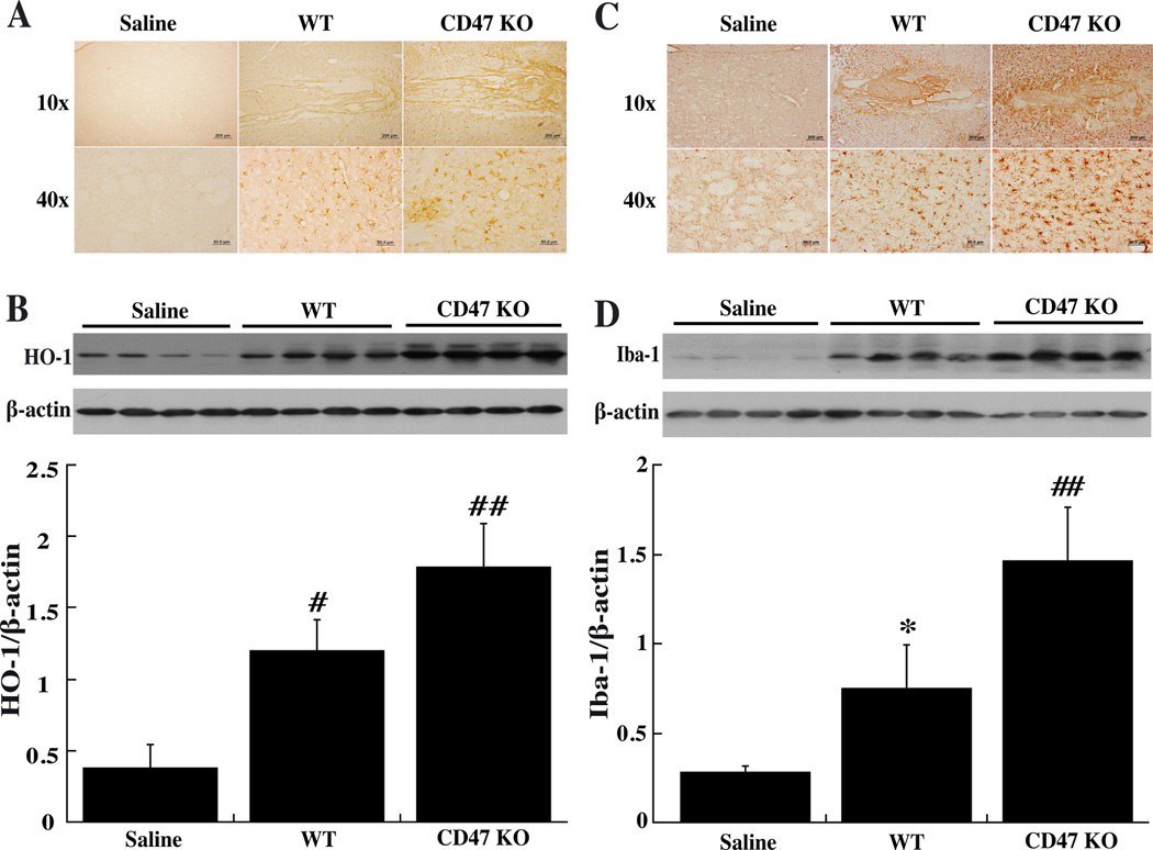
Figure 3.
Immunoreactivity and protein levels of HO-1 (A, B) and Iba-1 (C, D) in the ipsilateral basal ganglia at day-3 after injected of 30µl saline or blood from either WT or CD47 KO mice into the right caudate. Values are mean ± SD; n=4 for each group, *p<0.05, #p<0.01 vs. saline group; ##p<0.01 vs. the other groups.

