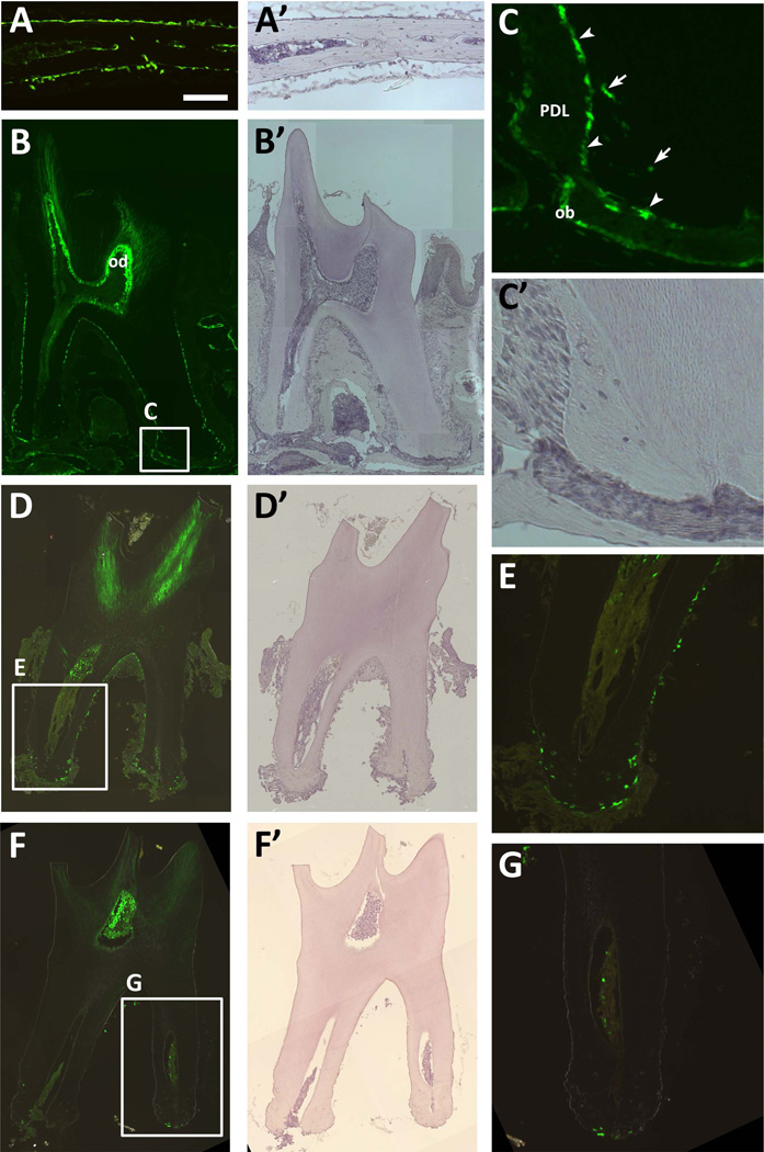
Figure 1. OC-GFP labels osteoblasts and cementoblasts.
(A) Expression of OC-GFP is restricted to osteoblasts in the calvaria. (B–C) A section of the second molar indicates that OC-GFP is present in cementoblasts (arrowheads) and cementocytes (arrows), as well as odontoblasts (od) and osteoblasts (ob) lining the alveolar bone, but not cells within the periodontal ligament (PDL). (D–E) After tooth extraction, cementoblasts remain on the tooth surface, but osteoblasts associated with alveolar bone are absent. (F–G) Following digestion, cells are no longer present on the surface of the tooth, including OC-GFP+ cementoblasts. A 100µm scale bar for the higher magnification images (A, C, E, G) is shown in A. A’-D’ and F’ show hematoxylin staining for the corresponding fluorescent image.

