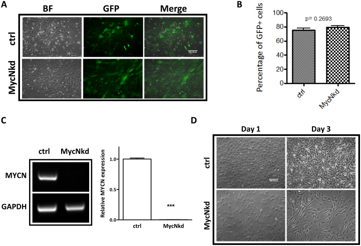
Fig 3. MycN knockdown inhibits cell growth in hNCSCs.
FACS-sorted p75+ hNCSCs were plated at a cell density of 4×10³ cells/cm2 in self-renewal media on 6-well plates that were pre-coated with 15 μg/ml polyornithine, 1 μg/ml laminin and 10 μg/ml fibronectin for 24 hours. Freshly isolated hNCSCs were transduced with concentrated pGLVH1/GFP-MycNshRNA virus and selected in puromycin (2 μg/ml) for two weeks, as described in Materials and Methods.(A): The GFP expression in control and MycN-transduced hNCSCs; (B): FACS analysis shows the similar transduction efficiency in control and MycN-transduced hNCSCs; (C): RT-PCR analysis showed the expression of MycN in control and MycNkd hNCSCs after 2 weeks of antibiotics selection. Image shown is the representative of three independent experiments; (D): Freshly sorted cells were plated onto 15 μg/ml polyornithine, 1 μg/ml laminin and 10 μg/ml fibronectin coated plates at a concentration of 10x103 cells/well and grown in NC media. Within two days to adherent plate, MycN knocked down cells initiated a change from small, NCSC-like to a larger shape.

