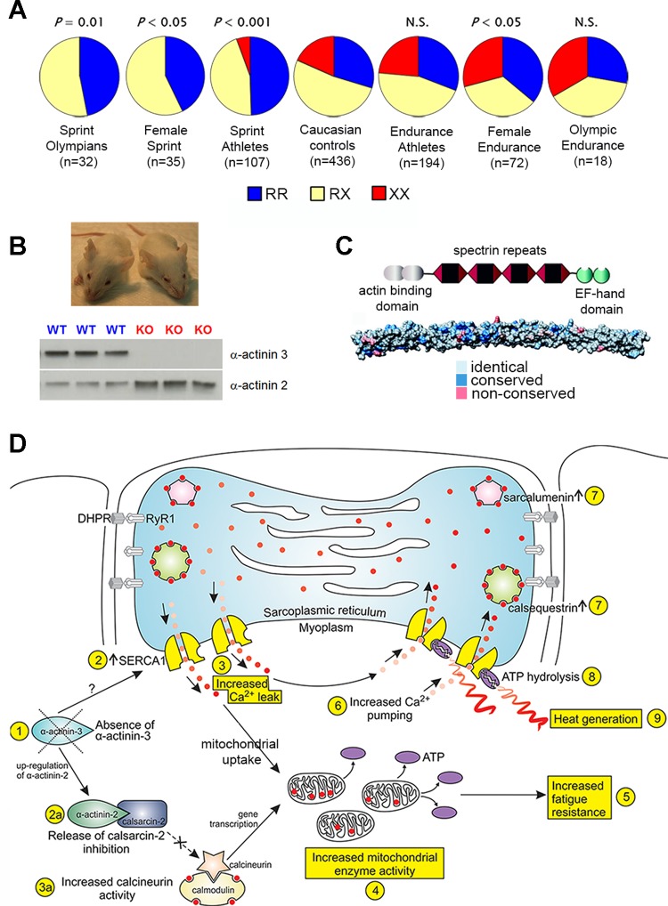
Fig. 5.
A: the frequency of R577X ACTN3 genotypes (%RR, RX, and XX) differs in sprint populations (lower frequency of the XX genotype) compared with controls and endurance populations (higher frequency of the XX genotype in females) [data adapted from MacArthur et al. 2008 (51)]. B: WT and Actn3 knockout (KO) mice (pictured respectively) have been indispensable in modelling the human phenotype. Actn3 KO mice do not express α-actinin-3 on Western blot and have an upregulation of α-actinin-2 in skeletal muscle [MacArthur et al. 2008 (51)]. C: the highly conserved domain structures of α-actinin, with an in silico surface representation of the rod domain. The colors represent identical, conservative, and nonconservative residue substitutions when α-actinin-2 and -3 vertebrate sequences are aligned [figure from Lek et al. 2010 (46) used with permission from Elsevier]. D: in vitro and in vivo evidence demonstrates that the loss of α-actinin-3 alters calcineurin signaling, mitochondrial enzyme activity, and calcium handling (increased pump and release rates) to explain their improved fatigue resistance and also may result in enhanced heat production [figure from Head et al. 2015 (33)].

