
Fig. 11.
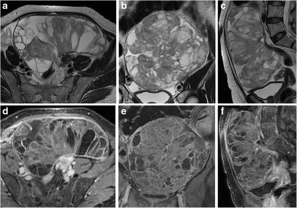
Mucinous cystadenocarcinoma in a 57-year-old woman. (a) Axial, (b) coronal and (c) sagittal T2-weighted images show a large mass with mixed solid and multilocular cystic appearance with low signal intensity of the solid component and variable signal intensity within the locules (“stained glass appearance”). (d) Axial, (e) coronal and (f) sagittal contrast-enhanced fat-suppressed T1-weighted images demonstrate marked enhancement of the solid component, wall and septa of the tumour