
Fig. 16.
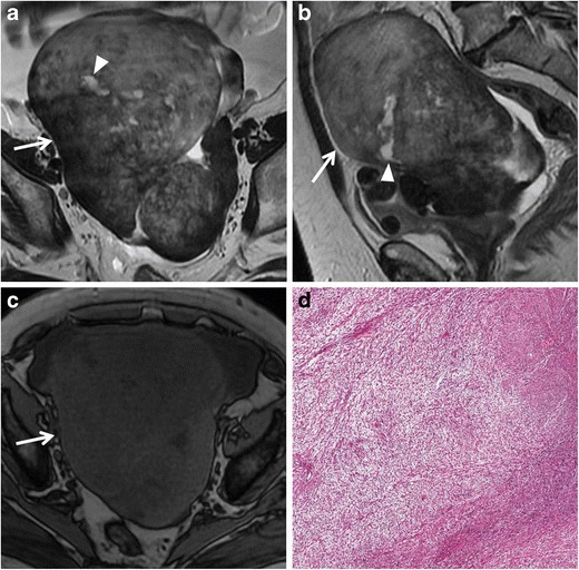
Fibrothecoma in a 66-year-old woman. (a) Axial and (b) sagittal T2-weighted images show a heterogeneous solid mass (white arrows) with low to intermediate signal intensity. Some areas of cystic degeneration with high signal intensity can be seen within the lesion (white arrowheads). (c) Axial T1-weighted image confirms the solid appearance of the lesion (white arrow). (d) Photomicrograph (H&E X200) shows fascicles of spindle cells with centrally placed nuclei and a moderate amount of pale cytoplasm