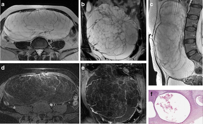
Fig. 4.

Mucinous cystadenoma in a 44-year-old woman. (a) Axial, (b) coronal and (c) sagittal T2-weighted images show a voluminous multilocular cystic mass with several septations. (d) Axial and (e) coronal contrast-enhanced fat-suppressed T1-weighted images demonstrate poor enhancement of the tumour wall and septa. (f) Photomicrograph (H&E X400) shows cystic spaces lined by columnar cells of intestinal type with mucinous secretion
