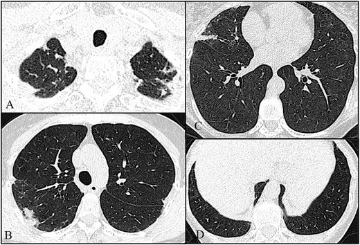
Fig. 4.

Axial high-resolution chest tomography (HRCT) images (a–d) of patient 4 show pleural and subpleural thickening with moderate fibrotic changes in the marginal parenchyma with apical-caudal distribution. These features are more evident in the right lobe. Note also, in c, a slight “tree-in-bud” pattern in the middle lobe compatible with concomitant pulmonary infection
