
Fig. 4.
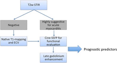
Revised diagnostic algorithm for the clinical workup in patients with clinically suspected acute myocarditis. Routine inclusion of T1 mapping techniques (native and ECV) in the scanning protocol would enable the coupling of the high specificity of T2-STIR and LGE techniques with the increased sensitivity of T1-relaxation changes measurements (particularly in mild focal or diffuse forms of disease). According to the literature, a combination of functional data and inflammation/necrosis imaging correlates provided by CMR may serve as a predictor of functional and clinical recovery at follow-up (see text for further explanation).