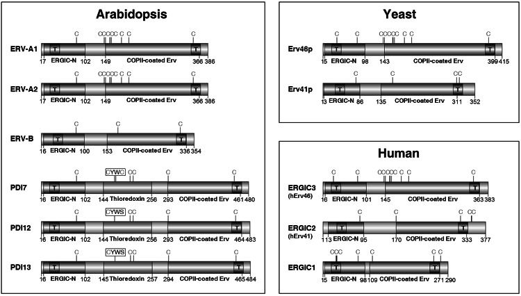
Fig. 2.
Domain arrangements of Erv41p/Erv46p proteins from Arabidopsis, yeast, and humans. ERGIC-N domains are shown in blue, COPII-coated ERV domains in green, and thioredoxin-like domains in red. Black boxes labeled with the letter T indicate the predicted positions of TMDs. The positions of Cys residues are indicated by the letter C (colour figure online)

