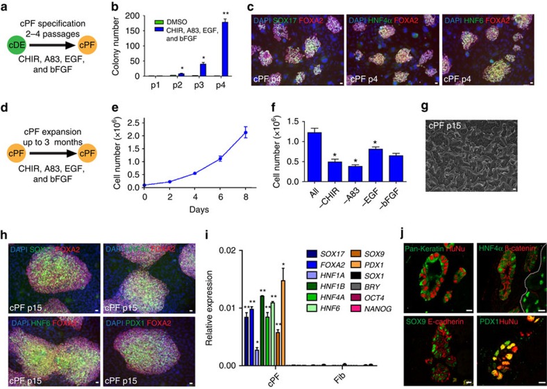
Figure 2. Specification, expansion and characterization of posterior foregut-like progenitor cells.
(a) Schematic representation of culture conditions for the amplification of cDE cells that resulted in further specification into expandable posterior foregut-like progenitor cells (cPF cells). (b) Improved culture conditions allow amplification of cDE/cPF cell colonies. Mean values±s.e.m. represent three experiments. Statistical significance calculated using two-tailed Student's t-test, compared with DMSO controls. *P<0.05, **P<0.01. (c) Immunofluorescence analysis of colonies after four passages in improved expansion media suggested specification towards cPF cells. Scale bar, 20 μm. (d) Illustration of cPF expansion strategy. (e) Growth curve of cPF cells. Data from three experiments are shown as average±s.e.m. (f) All four media supplements—EGF, bFGF, A83-01 and CHIR99021—are important for cPF cell self-renewal (n=3 experiments). Statistical significance calculated using two-tailed Student's t-test, compared with ALL supplementation. *P<0.05. (g) Bright-field image of established cPF cells showing epithelial colony morphology. Scale bar, 20 μm. (h) Immunofluorescence staining of SOX17, FOXA2, HNF4α, HNF6 and PDX1 in cPF cells at passage 15. Scale bar, 20 μm. (i) QPCR analysis demonstrates the enrichment of transcripts for SOX17, FOXA2, HNF1A, HNF1B, HNF4A, HNF6, SOX9 and PDX1, but not SOX1, BRY, OCT4 and NANOG in p15 cPF cells. Mean values±s.e.m. are normalized to GAPDH relative to control fibroblasts. (n=3 experiments). Statistical significance calculated using two-tailed Student's t-test, compared with fibroblast controls. *P<0.05, **P<0.01. (j) Immunofluorescence analysis of cPF cell grafts shows epithelial structures that express E-cadherin, HNF4α, PDX1, SOX9 and pan-cytokeratin. Human nuclear antigen (HuNu) demonstrates the human cell origin. Scale bar, 20 μm.

