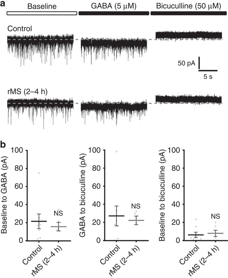
Figure 2. Tonic GABAAR conductance is not affected after rMS.
(a) Sample traces depicting GABA- (5 μM) and bicuculline-methiodide (50 μM)-induced shift in tonic GABAAR-mediated currents recorded from CA1 pyramidal neurons in non-stimulated control and stimulated slice cultures (2–4 h after rMS). Baseline currents were not significantly different between the two groups (control: 191.2±12.1 pA; rMS: 187.5±13.6 pA; P=0.57; control, n=8 neurons; rMS, n=6 neurons; one cell per culture; Mann–Whitney test). (b) The amplitude of GABA- or bicuculline-methiodide-induced shifts in tonic GABAAR currents of CA1 pyramidal neurons is not significantly different between stimulated and non-stimulated slice cultures (control, n=8 neurons; rMS, n=6 neurons; one cell per culture; Mann–Whitney test). Individual data points are indicated by grey dots. Values represent mean±s.e.m. (ns, not significant differences).

