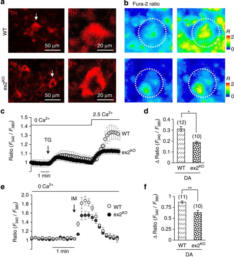
Figure 3. Store-operated Ca2+ signalling in iPSC-derived DA neurons from WT and ex2KO mice.
(a) Representative images of iPSC-derived A9 midbrain DA neurons positive for TH (TH+) from WT and PLA2g6 ex2KO mice (see Supplementary Fig. 12 for more details); (b) images demonstrate Ca2+ changes due to SOCE in specific DA neurons outlined by dotted circles and shown by an arrow in a: images show Fura-2 Ratio (F340/F380) in individual TH+ neuron before (left) and after (right) Ca2+ addition to TG-pretreated cells, as shown in c. (c) TG-induced SOCE (average±s.d.) in individual iPSC-derived DA (TH+) neurons from WT and ex2KO mice: traces show Ca2+ changes in response to TG (5 μM) application in the absence of extracellular Ca2+, followed by SOCE on Ca2+ addition. (d) Summary data comparing the peak SOCE (average±s.e.) in DA (TH+) neurons from WT and ex2KO mice. (e) Ionomycin (IM; 100 nM)-induced Ca2+ release (average±s.d.) from intracellular stores in DA (TH+) neurons from WT and ex2KO mice. (f) Summary data from experiments like in f show peak Ca2+ release (average±s.e.). The data represent the results from three independent experiments. The numbers of cells analysed for each condition is specified above the bars; *P<0.05, **P<0.01.

