FIGURE 7.

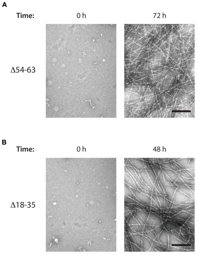
MccE492 mutants Δ18–35 and Δ54–63 preserve the ability of forming amyloid fibrils in vitro. Negative-stain electron microscopy visualization of amyloid fibers formed by the MccE492 variants Δ54–63 (A) or Δ18–35 (B), incubated in aggregation buffer until the indicated time. Scale bar: 100 nm.
