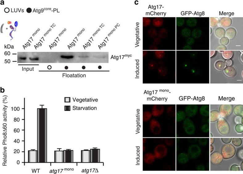
Figure 5. The dimerization domain of Atg17 regulates Atg17-function.
(a) α-myc immunoblots from co-floatation experiments of Atg9–PLs with recombinant myc-tagged, monomeric Atg17 (Atg17mono), Atg17monoTC and Atg1monoPC. Atg17mono is strongly inhibited by Atg31–Atg29, but Atg17monoTC is not activated by Atg1–Atg13. Similar experiments with large unilamellar vesicles (LUVs) lacking Atg9core served as controls. Input corresponds to 10% of total protein used for co-floatation. (b) Pho8Δ60 assay of wildtype (WT), atg17mono, and atg17Δ cell lysates to assess autophagic activity under vegetative (grey bars) and starvation (black bars) conditions. Mean values±s.d. of N=3 independent experiments are shown. (c) Co-localization of GFP-Atg8 with mCherry-tagged Atg17 or Atg17mono under vegetative or autophagy-induced conditions. Atg17mono is not forming puncta under autophagy-induced conditions. Consequently, autophagosomes are not being formed as apparent from the lack of Atg8-puncta. Scale bar, 2 μm.

