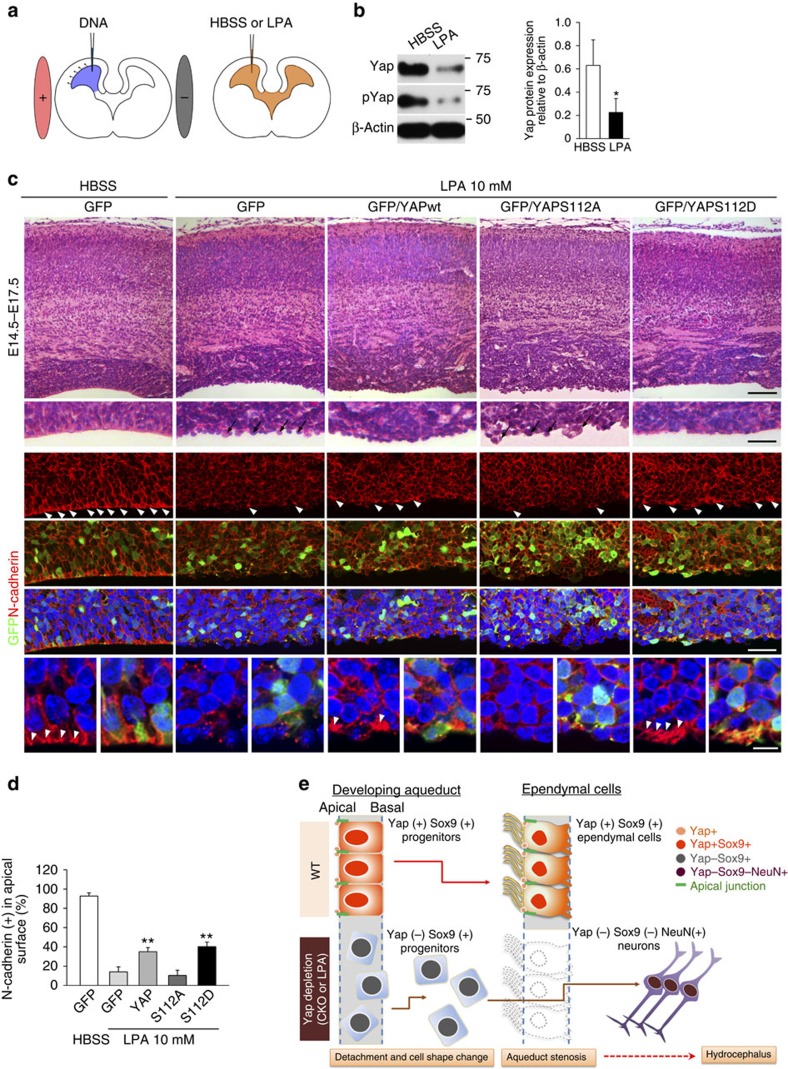
Figure 8. LPA-induced junction defects are partly rescued by forced expression of Yap.
(a) Schematic of the rescue experiments: electroporation of Yap or Yap mutant constructs to the developing cortices followed by LPA administration to a lateral ventricle. (b) Yap and pYap are significantly reduced in the cortex 1 day after LPA injection into lateral ventricle at E13.5. β-actin was used as a loading control. (c) Histological analysis of an electroporated area of cortex after LPA reveals disruption of the ventricular surface: the apical lining of cells is segregated and lacks lateral attachment to adjacent cells. Number of protruding cells in the apical surface is reduced by Yap overexpression and more prominently by phosphomimetic Yap (S112D) expression. Phospho-defective Yap (S112A) expression fails to reduce these protruding apical surface cells, but expansion of VZ is evident. N-Cadherin localization at the cell junction and apical surface (arrow heads) is markedly decreased by LPA as compared with control (HBSS treated), and is partially restored by forced expression of Yap and phosphomimetic Yap (S112D), but not by phospho-defective Yap (S112A). (d) Number of N-Cadherin (+) cells at the apical surface counted and compared. (e) Schematic showing that loss of Yap by genetic deletion or LPA treatment results in disrupted junction proteins and a failure to form the ependymal layer. Two-tailed unpaired t-test reveals statistical significance (**P<0.01 and *P<0.05). Error bars represent mean+s.e.m.; n=3. Scale bars, 100, 40 and 10 μm (c).

