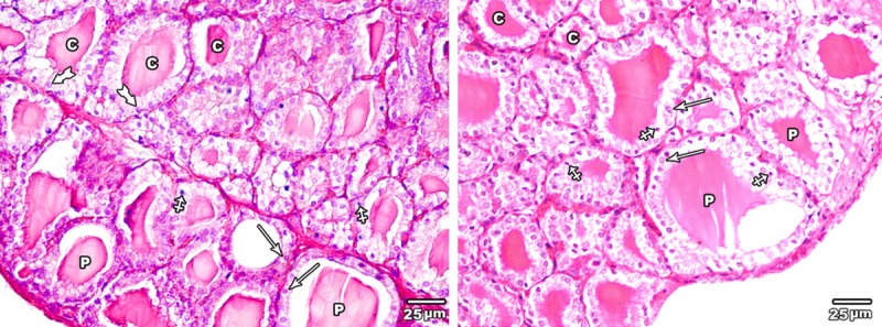
Figure 5.

A section in the thyroid gland of adult rat treated with low dose of MSG (group II) the thyroid follicles showing large peripheral follicles (P) lined with cubical epithelium with central rounded nuclei (arrow) and central ones (C) lined with columnar epithelium with basal nuclei (tailed arrow). Note few pyknotic nuclei (crossed arrow) were seen (H&E × 400).
